Chilblains
Last edited on : 22/10/2023
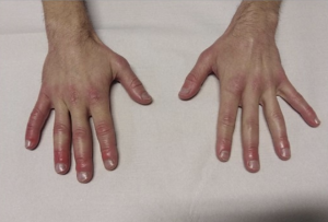
Chilblains (or erythema pernio) is a paroxysmal vascular acrosyndrome characterized by the appearance of pruritic, erythrocyanotic lesions on the extremities, reflecting ischemia due to arteriovenous vasospasm following prolonged exposure to moderate cold and arteriolar vasodilatation with venous spasm persisting on reheating. The prevalence of the condition is high (around 2.5% in women and 0.5% in men). It should be distinguished from frostbite (ischemic lesions resulting from prolonged exposure to intense cold).
This condition is idiopathic and generally benign. Atypical forms, however, require further work-up to exclude differential diagnoses (pseudo-chilblains).
Clinic

Positive diagnosis is essentially clinical. Typical forms :
- Occurs in young or adolescent women, thin in winter.
- Triggering factors: shoes that are too tight, insufficient protection against the cold, poorly insulated or poorly heated accommodation, medication (vasoconstrictors, beta-blockers, etc.).
- Appearance of lesions on warming, usually consisting of one or more painful, itchy, erythrocyanotic papules confluent into infiltrated plaques with poorly-defined contours. Other, rarer forms are possible: lenticular, cocarde, keratotic, bullous or ulcerated lesions. Recurrences are frequent (2 to 3 times per winter), generally in the same locations.
- Preferentially affecting toes and fingers, but sometimes heels, nose, knees. Single or bilateral.
- Peripheral pulses are normal on clinical examination (if not, suggest obliterative arteriopathy).
Lesions usually regress spontaneously and without complication within a few weeks. More rarely, they may become superinfected, ulcerated or necrotic.
The condition may also be associated with another acrosyndrome (++ acrocyanosis and Raynaud's phenomenon).
Differential diagnosis
There are many pathologies likely to manifest as "pseudoengelures", and these should only be considered in cases of clinical atypia (male patient, patient > 50 years of age, ulcerations, atypical localizations, occurrence outside a winter context, semiology or context suggestive of another pathology, abnormal peripheral pulses, etc.):
- Erythema multiforme
- Systemic lupus erythematosus
- Cold-related dermatoses (panniculitis, urticaria, etc.)
- Vasculitis
- Atheromatous arteriopathies
- Platelet or cholesterol embolisms
- Other vascular acrosyndromes
- Malignant hemopathies, paraneoplastic syndromes, multiple myeloma, cryoglobulinemia
- Miscellaneous: scleroderma, sarcoidosis, tertiary syphilis, jellies, compartment syndrome, various infections and post-vaccination reactions, etc.
Additional tests
Typical benign forms do not require further investigation. However, some authors recommend systematic capillaroscopy and laboratory tests (hemato-VS-CRP, FAN, cold agglutinins, cryoglobulin). In all cases, a more extensive work-up is required only in the event of clinical atypia, in order to exclude differential diagnoses.
- Capillaroscopy and pulp doppler
- Biology: hemato-VS-CRP, FAN, cold agglutinins, cryoglobulin +- cryofibrinogen, anticoagulant antibodies, rheumatoid factor
- Arterial Doppler ultrasound
- Biopsy of lesions
Therapeutic management - Treatments
- Avoidance of and protection from cold and damp, cessation of smoking, avoidance of drugs and toxic substances (vasoconstrictors and beta-blockers), correction of any dietary disorders.
- In the case of incapacitating forms of the disease: anticalciques in crisis (e.g. diltiazem), fatty creams.
- In case of ulceration or necrosis: assessment by a vascular surgeon, hemodilution, anticalciques
- Management of any associated acrosyndrome
Author(s)
Dr Shanan Khairi, MD
Bibliography
EMC, Traité d'Angéiologie, Elsevier, 2014
EMC, Traité de médecine, Elsevier, 2009
Creager M et al., Vascular Medicine: A Companion to Braunwald’s Heart Disease, Saunders, 2013
Longo DL et al., Harrison - Principes de médecine interne, 18e éd., Lavoisier, 2013
Pileri A et al., Chilblain lesions after COVID‐19 mRNA vaccine, British Journal of Dermatology, Volume 185, Issue 1, 1 July 2021